The Importance of Dental X-rays
Dental x-rays (or radiographs) are critical for diagnosing disease under the gumline. 60% of the tooth anatomy cannot be seen, so without these x-rays, the disease can be left undiagnosed. If left untreated, dental disease can not only cause serious pain and loss of appetite, but it can also lead to long term health problems such as heart and kidney disease or serious problems like a jaw fracture.
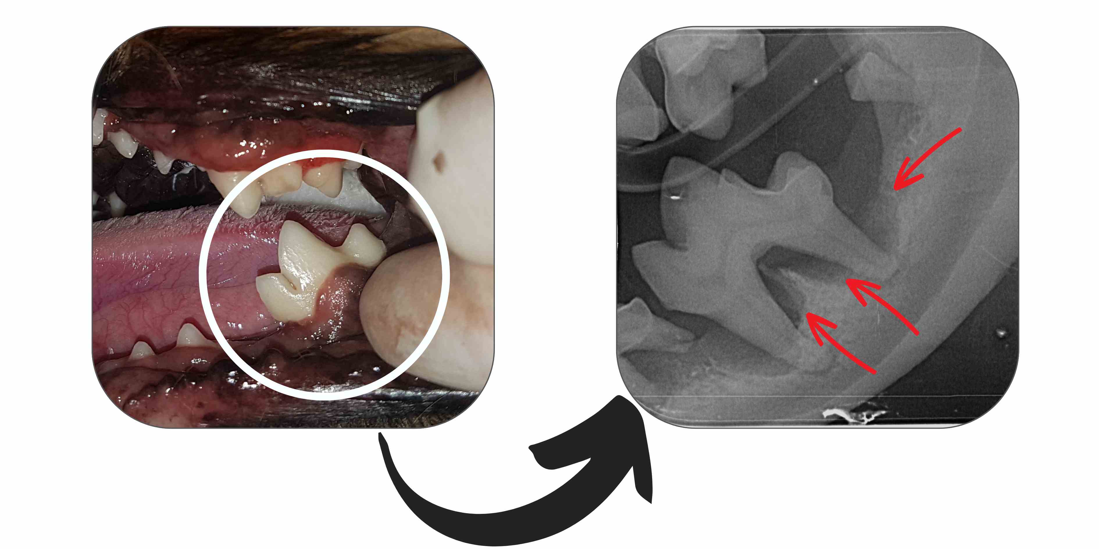
The image above depicts a seemingly normal tooth. However, upon examination with dental X-rays, severe bone loss or disease surrounding the tooth becomes visible, as indicated by the red arrows.
Dental x-ray helps us be better prepared for extractions or surgical procedures. It also lets us know when a procedure is highly complicated and needs to be referred to a dental specialist.
In most cases, dental x-ray is recommended at every dental procedure, both before and after the procedure. The initial x-rays also provide a reference of the teeth's health for future comparison as your pet ages.
Dental radiography assesses 3 main areas of dental health:
1. Internal anatomy of the teeth
2. Dental pathology
3. Treatment success
Conditions That Can Be Diagnosed
Common, often painful, pathology that may not be visualised externally can be diagnosed with x-rays, therefore enabling your Veterinarian to formulate a treatment plan to rectify the problem.
These conditions include:
- Retained deciduous teeth (baby teeth that failed to shed at the proper time)
- Missing teeth
- Unerupted or impacted teeth
- Tooth fractures
- Resorptive lesions (common in cats)
- Crowding and rotation of teeth
- Periodontal disease
- Jaw problems e.g. malocclusions, jaw fractures, cysts
- Oral growths
- Tooth root abscesses or infections
- Bone or soft tissue tumours
- Height of the bone below the gum line
Dental Disease Classification
Radiographs can help classify periodontitis (dental disease):
Mild - if < 25% of alveolar bone has been lost
Moderate - if 25-50% of alveolar bone has been lost
Severe - if >50% of alveolar bone has been lost
This classification is essential in determining the best treatment plan and prognosis for each tooth. Depending on the level of periodontitis, the treatment may include extraction, periodontal surgery, or conservative management.
Post-Treatment Radiographs
After dental treatments such as extractions, post-treatment radiographs play a crucial role in evaluating treatment success.
Tooth roots, often curved (or hooked) in structure, can pose challenges during extraction. Identifying its presence prior to extraction allows the Veterinarian to successfully extract the root with greater precision, minimising complications.
Studies indicate that nearly 90% of oral surgery cases can have root fragments remaining in the socket after extraction, which can cause future complications if left behind. Radiographs are indispensable for detecting any these root fragments and guiding the Veterinarian to perform complete extractions.

In the first image above on the left, the red arrows highlight the curved root (hooked appearance at the root tip), indicating its presence. In the second image on the right, marked with a white circle, you can observe the retained tooth root.