
A Guide to Pet Lumps and Bumps
Finding lumps and bumps is a common problem seen in middle-aged to senior dogs. It can be a very distressing find, but fortunately, most small skin lumps found in dogs can be cured when removed surgically. Early detection and ongoing monitoring is the key to successful treatment.
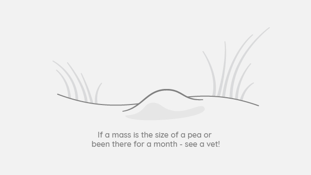
The overall recommendation for lumps is that if the skin lump is greater than the size of a pea or has been there for over a month, it should be further investigated and surgically removed. Lumps that are left can grow too big or have the opportunity to spread locally or to other parts of the body.
Treatment is recommended immediately if the lump:
Bleeds
Changes colour
Changes shape
Is larger than pea-size
Grows quickly and suddenly
Causes pain
Types of Lumps
Lumps come in all shapes, types, sizes, locations, and behaviour (benign or malignant). All these factors play an important role in determining the best course of action.
Malignant tumours are those that are aggressive and spread locally or to other parts of the body. Of all the skin tumours, it is estimated that 20-40% of them will be malignant in dogs and as high as 50-70% will be malignant in cats.
On the other hand, benign tumours have a better prognosis and do not spread. For example, fatty lumps or lipomas do not pose any harm to a pet and do not require any treatment if it does not grow quickly or interfere with limb movement.
Even to the most trained eye, a diagnosis cannot be accurately made on the appearance of the lump alone. Lumps of varying origins and diagnoses (benign or malignant) can have very similar physical appearances. Further tests such as a biopsy or fine needle aspirate are required to collect the lump cells and tissue for analysis to determine the type of tumour and the best treatment plan.
Preliminary Testing
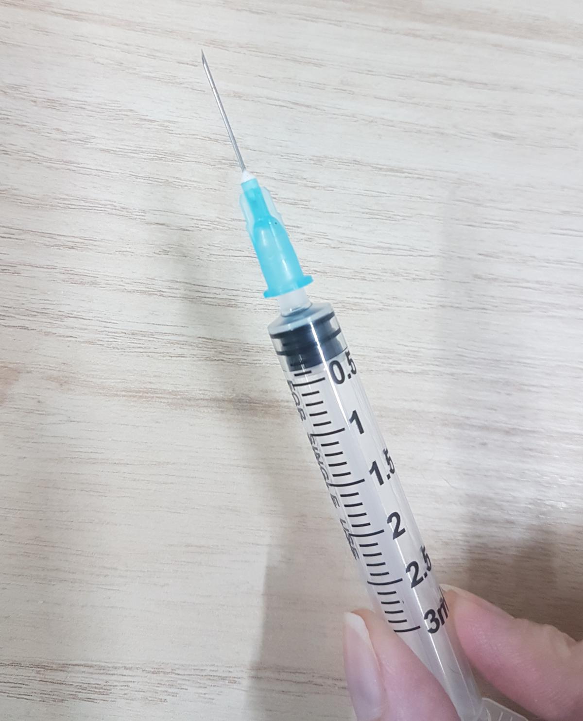
Fine needle aspiration (FNA) is the most frequently used technique in cytology. It is typically used to sample “lumps and bumps” on the body. This technique can also be used to evaluate internal organs, such as the liver, lungs, lymph nodes, or kidneys and body fluids, such as urine or joint fluid.
During an FNA, a very thin needle is used to collect a sample of cells, tissue or fluid from an abnormal area or lump. In some cases, an ultrasound may be required to obtain accurate sampling by FNA.
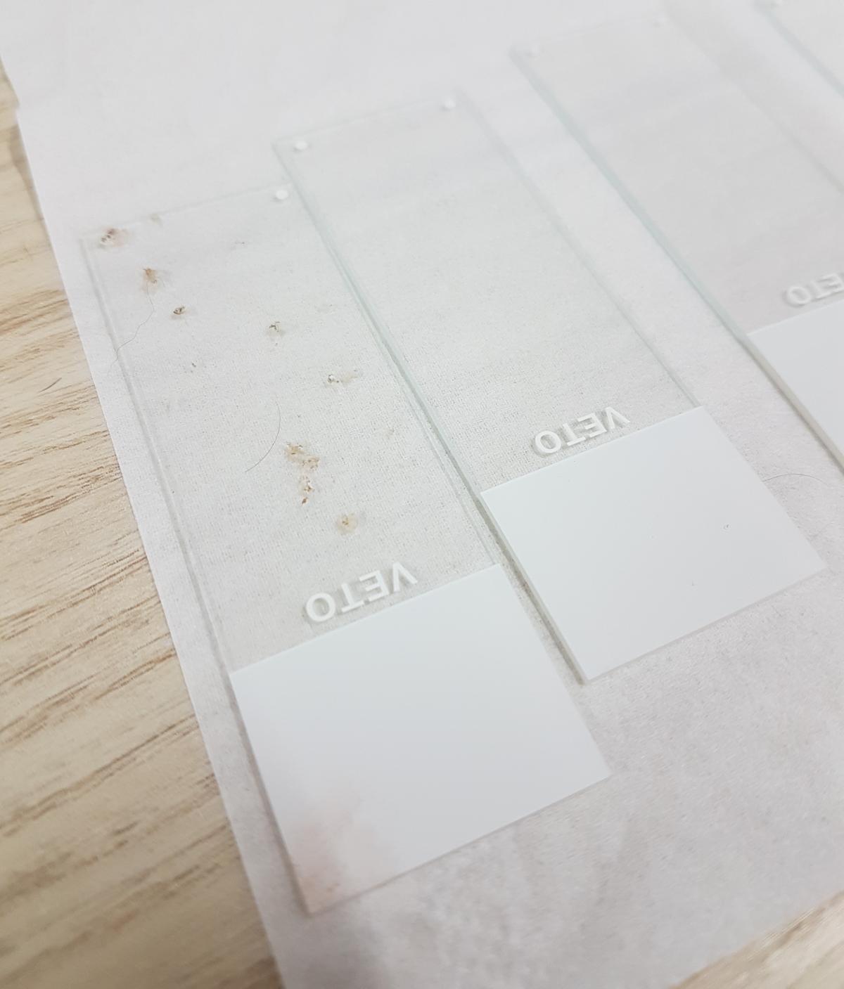
The sample is carefully placed on to glass slides to be sent to a reference laboratory for microscope analysis. FNAs can yield a diagnosis in 70% of cases. For the other 30% deemed as “non-diagnostic”, possible reasons may include lumps that do not exfoliate cells or tissues readily, leading to inadequate tissue representation.
Most patients will tolerate an FNA without the need for sedation. However, anxious patients, or lumps located in sensitive areas such as the face, armpit or groin, may require heavy sedation or even a general anaesthetic.
The result of an FNA enables your Veterinarian to determine the margins of excision (how much tissue around the lump needs to be removed along with the lump). Benign lumps require minimal margins whereas malignant lumps often require very large margins.
Surgical Treatment
The goals of surgery are to:
Surgically remove the lump to reach a cure; or
Remove all visible lumps; or
Identify the mass to determine a treatment plan; or
Form part of a multimodal approach along with radiation or chemotherapy.
Surgery is performed in a sterile manner that helps to protect the normal tissues from tumour cell contamination. We use large surgical drapes, lavage, gloves and instrumental changes where required. The mass is minimally handled to reduce trauma.
Where possible, the lump will be removed in its entirety with clean margins both wide (at least 2 cm margins around the mass) and deep where required.
In cases where this cannot be achieved, the majority of the lump will be removed and samples sent away for pathology to determine the next steps. When suspicious of local lymph node involvement, a lymph node biopsy will be recommended.
Histopathology
After a lump is excised (removed from the body), it is recommended to have it sent to the reference laboratory for histopathology. This type of testing is where the lump is first preserved in formalin, then sectioned into microscopic thin layers and stained. These tissue sections are examined under the microscope by a veterinary pathologist. Histopathology focuses on the architecture of the tissue and provides more information about the tissue than cytology and the accuracy of a diagnosis is usually high.
Histopathology assists with:
Diagnosis of the tumour type and degree of malignancy
Determining if a complete excision has been achieved (i.e. no tumour cells extending past the cut edges)

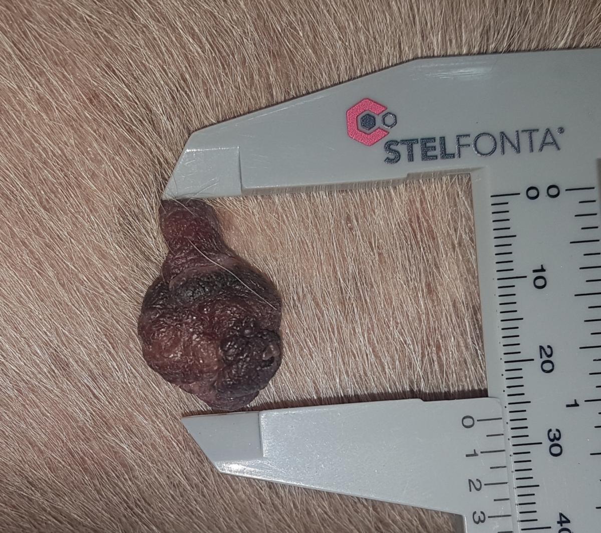
A lump to be sent to the reference laboratory for histopathology.