
Cruciate Ligament Repairs
Prognosis for dogs that undergo surgical repair is good with improvement seen in 85-90% of cases.
Overview
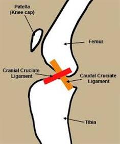
There are two bands of fibrous tissue called the cruciate ligaments in each knee joint. They connect the femur (the leg bone above the knee joint) and tibia (the leg bone below the knee joint) togethe
They are called cruciate ligaments because they "cross over" inside the knee joint. The cranial cruciate ligament limits the forward movement of the tibia in relation to the femur. It also limits hyperextension (over straightening) of the knee and internal rotation (turning in) of the tibia.
Cruciate ligament rupture is a common knee injury of human athletes (the ACL that football players commonly injure). These "acute" or sudden ruptures can occur in dogs, but they are much less common than a process called "Cruciate Ligament Disease". This is the name given to the complex of issues that occur in the knee as a result of cruciate injury.
How does cruciate ligament disease occur?
The knee joint is a hinged joint and only moves in one plane; backwards and forwards. Cruciate ligament damage is caused by a twisting action to the knee joint. This injury usually affects the anterior or cranial (front) ligament. The knee joint then becomes unstable and causes pain, often resulting in lameness.
Why does cruciate ligament disease occur?
In dogs, a chronic form of cruciate damage occurs due to weakening of the ligaments as a result of disease. The reasons for this are only partially understood. The ligament may become stretched or partially torn during exercise, and lameness may only be slight and intermittent. In dogs, the top of the tibia is more slanted than in people, placing additional strain on the cruciate ligaments.
This begins a process of inflammation, or arthritis, in the joint. With continued use of the joint, the condition gradually gets worse until complete rupture of the ligament occurs in the course of normal activity. Some patients may suddenly yelp during physical activity and then become acutely lame. The activity that ruptures the ligament does not need to be violent nor excessive. 90% of cruciate ruptures in dogs occur in this way. Because this is a degenerative process, the knee joint will usually be arthritic before the cruciate disease is diagnosed. Additionally, the other knee joint usually follows suit in this progression of disease.
Clinical signs of a cruciate ligament rupture include lameness with very little weight-bearing. An injured dog usually holds the limb up or may only be intermittently lame. The treatment for this is typically surgery.
Occasionally, the cartilage within the knee joint called the meniscus can become damaged. The meniscus acts as a shock absorber between the leg bones and can cause extreme discomfort when damaged.
Signs
Common signs of a cruciate ligament rupture:
- Sudden, severe limping on one rear leg
- No or little weight bearing on the leg after an injury
- Mild or intermittent limping in the case of a partial tear
- Swelling of the knee may occur
- Difficulty rising or jumping
Predispositions
Predisposition to cruciate ligament rupture:
- Large and giant breeds are at a higher risk than small breeds
- Young, active dogs are at higher risk
- Overweight dogs suffer higher levels of stress on their joints
- Dogs that have been hit by cars, attacked by other dogs or suffered other forms of trauma
- Dogs that have previously injured a cruciate ligament in one knee are at an increased risk of injuring the ligament in the other knee at a later date
- Dogs with relatively long legs
Diagnosis
A careful review of the patient's history and a complete physical examination is the first step. Many pets may "toe touch" and place only a small amount of weight on the injured leg. Your veterinarian may perform stifle manipulations such as the "cranial drawer" or "tibial thrust" test to determine the degree of joint laxity. Other diagnostic tests such as xrays are usually necessary.
Once the cruciate ligament is ruptured, the knee joint becomes unstable, acutely inflamed and painful. This instability can result in damage of the medial meniscus (a C-shaped piece of shock-absorbing cartilage located inside the joint). This is very commonly damaged and is a source of extreme pain.
We can often feel or hear a "click" in the knee joint of dogs with meniscal damage. If left unmanaged, the knee joint becomes thickened and chronically painful. The leg muscles may waste due to disuse.
Management
Treatment of a cruciate ligament rupture may consist of:
- Exercise restriction and cage rest for several weeks. This may be sufficient in small dogs and cats./li>
- Weight loss.
- Surgical stabilisation.
- Medical management of osteoarthritis.
Dogs under 10kg may improve without surgery, especially aged patients. These patients are often restricted to cage rest for four to six weeks minimum. Dogs over 10kg usually require surgery to heal.
Unfortunately, most dogs (small or large) will eventually require surgery to correct this painful problem. It is best to perform surgery early on in the disease process for the best outcome.
Prognosis
Unfortunately, the prognosis for ARDS is often poor, especially when they are not subjected to immediate and intensive veterinary treatment. This distressing condition has a typical mortality rate of around 50% in humans and is close to 100% in veterinary patients with no medical intervention. Although few reports describe successful recovery from ARDS, recovery is not impossible with intense critical and nursing care.
Surgery
Surgery for cruciate ligament disease aims to return patients to pain-free function and slow the progression of degenerative joint disease (arthritis). There are a number of surgical techniques used to repair the cruciate ligament and/or meniscus. Your veterinarian will determine the appropriate surgical technique based on the size and age of the dog and the degree of damage.
Traditional Techniques
These procedures aim to replace the function of the cruciate ligament and stabilise the knee joint.
- The extra-capsular technique involves placing a synthetic ligament on the outside of the joint capsule to "replace" the missing cruciate ligament. Some available techniques are the De Angelis and ISO toggle techniques. The disadvantage of these techniques is the break-down or failure of the synthetic ligament before the knee joint can produce enough scar tissue to stabilise.
- The intra-capsular technique involves harvesting a strip of ligament from the leg and swinging it within the joins capsule to "replace" the missing cruciate ligament. We perform this technique here at Karrinyup Small Animal Hospital and have achieved good long-term outcomes for the affected leg. The disadvantage of this technique is the level of pain and discomfort immediately after the surgery.
Current Techniques
These procedures aim to produce dynamic stability of the knee joint, using the leg muscles to stabilise it or changing the angle of the tibial bone, without having to rely on the cruciate ligament. A few examples of current techniques are TPLO (Tibial Plateau Levelling Osteotomy) and MMP (Modified Macquet Procedure).
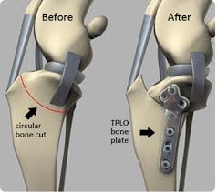
TPLO - Involves creating a circular cut in the top of the tibial bone and rotating the tibial plateau. This alters the dynamic of the knee joint to "stabilise" the knee when your dog walks. We have a travelling orthopaedic surgeon who comes to Karrinyup Small Animal Hospital to perform this procedure and has had great success rates. This is the preferred technique for large breed and highly energetic dogs.
Photo above: TPLO surgical technique
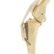
MMP - Involves creating a straight cut in the front of the tibial bone and advancing the bone forward. This creates a backward pull from the quadriceps muscle which indirectly supports the knee joint when weight is placed on the leg.
Photo above: MMP surgical technique
After Surgery
Your pet will remain in hospital for most of the day until it is fully recovered from the anesthetic and when its pain is under control.
Strict homecare following the procedure is extremely important to achieve the best outcomes. Excessive activity can result in poor healing or complications. It is important to follow the strict confinement regime to avoid surgical complications.
It is important that your dog has limited activity for six to eight weeks after surgery. Provided you are able to carry out your veterinarian's instructions, good function should return to the limb within three months. Unfortunately, regardless of the technique used to stabilise the knee joint, arthritis is likely to develop in the joint as your dog ages. To reduce the severity of arthritis, weight control, specific joint support diets, and joint support supplements should be commenced after surgery.
After surgery care may consist of:
- Crate or cage confinement for 10-14 days until the skin incision is healed.
- Your pet will have intradermal sutures (dissolving sutures in the skin). This means that your pet will require post-surgery checkups but no sutures will need to be removed.
- Room confinement and exercise restrictions for 8-12 weeks:
- No access to slippery flooring or stairs.
- No playing with other animals, running, jumping.
- Walking must be on a leash and controlled.
- Radiographs 8 weeks after surgery.
- Anti-inflammatory medications.
- Joint support injections eg Pentosan Polysulphate.
- Nutritional joint supplements eg 4-cyte or prescription joint diets.
If homecare instructions are not followed your pet may be subject to a number of complications after surgery. Potential complications may include:
- Soft tissue swelling
- Further meniscal injury
- Infection
- Implant failure
- Wound problem
- Patellar luxation (kneecap luxation)
- Failure to return to normal function
- Rapidly advancing arthritis
- Delayed bone healing, non-healing or incorrect bone positioning
- Bone fracture
What option is best for my dog?
After a careful physical examination of your dog, hip and hindlimb xrays and consideration of your home environment, your veterinarian will advise you on the best treatment option for your dog.
Rehabilitation
Studies show that physical therapy can improve the recovery process following this procedure. Your veterinarian may prescribe an exercise program specifically for your pet and advise you when to start.
Physical therapy examples:
- Passive range of motion
- Swimming
- Balance exercises
- Leash walking
Benefits of rehabilitation exercise:
- Help maintain good body condition
- Speeds recovery
- Prevents further injury
- Improves fitness and health
- Reduces the progression of osteoarthritis
- Improves mood
- Decreases pain
- Increases flexibility
- Improves blood flow
Prognosis
The prognosis for dogs that undergo surgical repair is good with improvement seen in 85-90% of cases. Surgery complications are uncommon but may include meniscal injury, infection, implant failure, and soft tissue swelling.
Although surgery can slow the progression of arthritis, it is still common for dogs to develop arthritis later in life.
Studies also show that approximately 50-60% of dogs will rupture the other cruciate ligament within 2 years of each other.
Although surgery is performed to minimise all complications, they can still happen from time to time, which may require further surgical intervention.
- Infection rates occur in 3-5% of patients
- Implant failures occur in 2% of patients
- Late meniscal injury occurs in 5-15% of patients
Prevention
Tips to help prevent a cruciate ligament rupture:
- Prevent obesity as this can lead to strained joints
- Provide regular exercise such as swimming to strengthen the muscles around the knee
- Stick to exercise that does not require a lot of twisting action or sudden stopping
- Always warm-up before strenuous exercise such as long hikes and running
A pet that is suspected of a cruciate ligament rupture should seek Veterinary attention immediately.
Why choose Karrinyup Small Animal Hospital?
At Karrinyup Small Animal Hospital, our cruciate surgery covers more than just the surgery itself; it also includes a comprehensive package of after-care, which is crucial to the success of this procedure.
This includes:
- Three days of post-op physical therapy (these are day stays, where your pet is dropped off to us in the morning, physical therapy is performed throughout the day and your pet is discharged in the afternoon).
- Veterinary rechecks at weekly intervals for six weeks, with Pentosan Polysulphate Injections (joint support injections) given at the rechecks between weeks 3 to 6 post-op.
- Post-op x-rays at 8 weeks to assess healing.
Your pet's wellbeing is very important to us. Our Team is skilled in understanding the numerous orthopaedic conditions your pet may face, and our hospital is designed for comfort and is a family-friendly environment.
Your pet's wellbeing is very important to us. Our Team is skilled in understanding the numerous orthopaedic conditions your pet may face, and our hospital is designed for comfort and is a family-friendly environment.
Contact us today to discuss your pet's Cruciate Surgery plan on (08) 9447 4644 or email [email protected]