
Linguoverted Teeth
Overview
Linguoverted teeth are a common dental concern seen in veterinary practice. It describes mandibular (lower jaw) canine teeth that are in the normal anatomical position but are angled abnormally, causing them to make contact with the teeth within the top jaw or hard palate tissue. This can result in trauma, discomfort and pain, and when left untreated can cause oronasal fistulas (canal between the mouth and nose). It can occur in deciduous or permanent teeth and is referred to as a Class 1 malocclusion of the mandibular canine tooth.
Predispositions:
Narrow and long-muzzled dogs
Large-sized breeds
Retained baby (deciduous) teeth
Hereditary
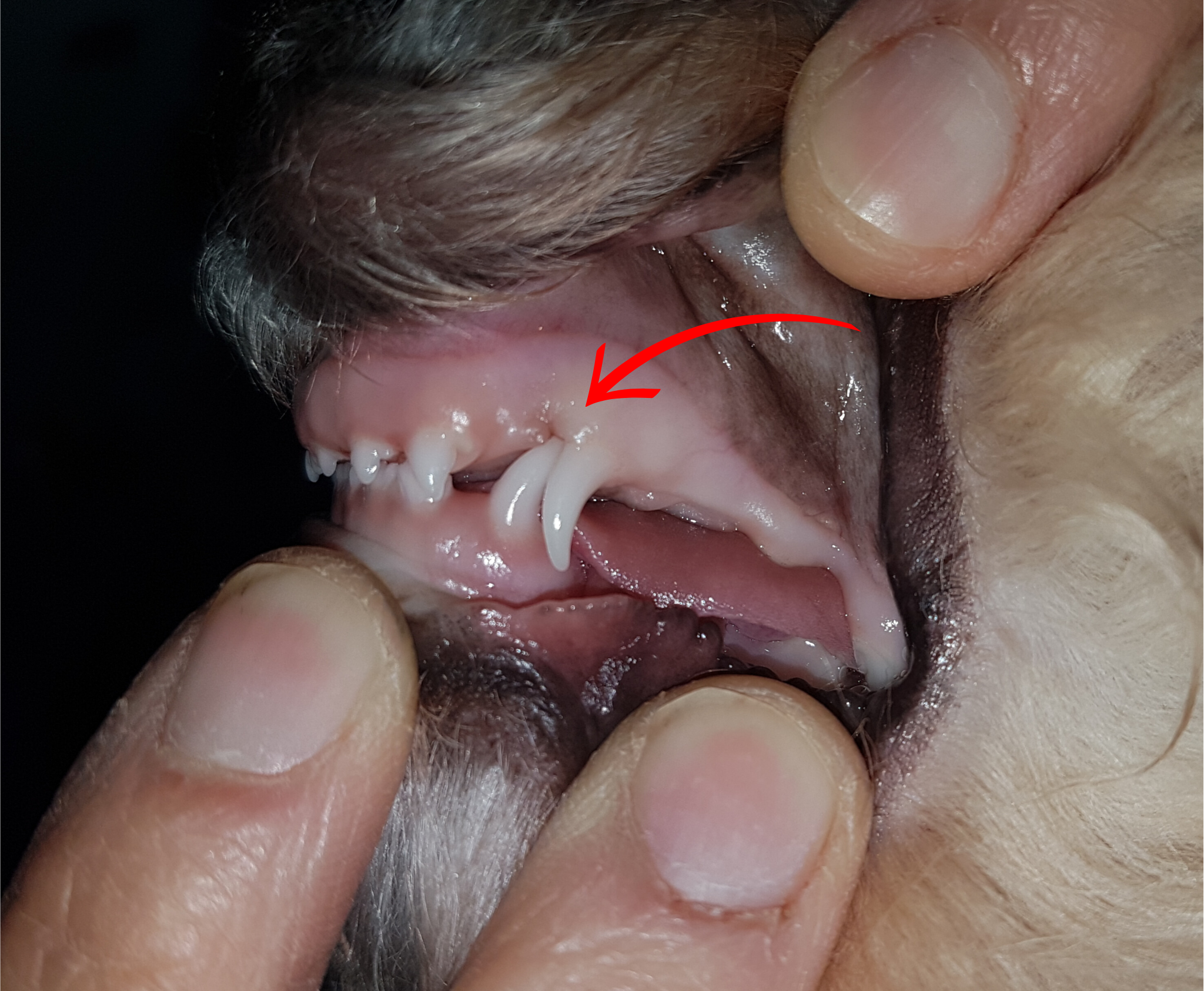
Photo showing an indentation in the hard palate caused by the linguoverted mandibular canine below.
.png)
Management
Your Veterinarian's recommendations will depend on your pet's lifestage and tools available to them. Referral to a veterinary dental specialist is also an option.
Extraction
When detected early enough (i.e. around 8-10 weeks of age), extraction of the linguoverted deciduous canine tooth is the recommended treatment and this procedure can be done here at Karrinyup Small Animal Hospital (KSAH). Risks of this procedure include general anaesthesia in a young pup and potential enamel damage to the budding adult canine tooth. As the aim is to create space for the adult tooth to grow out correctly, the benefit tends to outweigh the risks. Our experience at KSAH is that 90% of the time, early extraction results in the adult canine tooth growing out correctly and further intervention is not required.
However in some cases, the adult tooth may still develop a malocclusion. This is when the following options can be explored.
Ball Therapy
Should only be performed on adult teeth (NOT deciduous or baby teeth).
The ideal time to start this therapy is when the lower adult canine teeth are almost making contact with the hard palate. The window of opportunity for this intervention is very short i.e. around 6 weeks only.
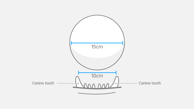
A study has shown that holding a rubber device e.g. spherical, hard, but hollow, rubber chew toy ball, for a minimum of 15 minutes, 3 times a day, may correct this malocclusion. The ball can help force the affected teeth to move into place as the dog bites down on the ball.
The appropriate size ball is imperative for success. The ball diameter should be around 1.5 times the distance between the 2 lower canines.
The Kong Classic dog toy is ideal for ball therapy. As it is also refillable (meaning you can insert treat food into it), it offers the dual-purpose of being both an enrichment tool, as well as helping to realign linguoverted teeth. The Kong Classic is available in three different sizes and our friendly staff can assist you in selecting the correct one for your pet. Tennis balls are NOT recommended, as they are too abrasive and will wear down the tooth's enamel.
In this study, 14 dogs underwent ball therapy for a Class 1 linguoversion malocclusion and their teeth were corrected between a period of 2 weeks to 2 months.
Crown Amputation
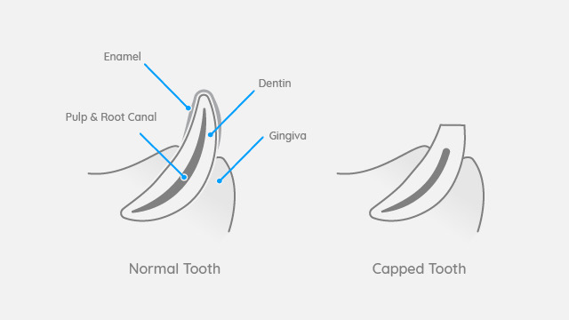
Another surgical procedure is a crown amputation and partial coronal pulpectomy on the adult canine. This involves surgically removing the tip of the lower canines and capping the tooth. The success rate is around 92% and the benefit of the procedure is that the tip of the canine no longer causes trauma to the hard palate or other teeth, and its root continues to maintain the strength of the lower jaw. Ongoing teeth radiographs are recommended 4-6 months after surgery and again 12-16 months. Failure tends to occur over 12 months post-operatively. The ideal candidate for this procedure is a dog that is NOT an avid chewer of hard solid objects such as rocks and sticks.
Full Canine Extraction
The full adult canine tooth can be surgically extracted, however, it is not an easy surgery (due to the long and large tooth root) and the void that is left can impact the stability of the lower jaw. The void is usually filled with a synthetic bone matrix to encourage bone regrowth for jaw strengthening.
Gingival Wedge
In mild cases, a wedge of gum can be removed from the top jaw to allow for space for the tooth.
Inclined Plane
The temporary application of a fixed acrylic or metal orthodontic device, placed under general anaesthesia to help direct the tooth into a healthier position, with correction within 2-4 weeks. This device must be removed once the tooth is corrected.
Camouflage Orthodontics
Tooth extensions that are made out of dental plastic also help change the direction of the tooth. When the proper position has been established, the plastic device is removed.
References
Wiggs, RB, Lobprise, HB. Basics of orthodontics. In: Wiggs, RB, Lobprise, HB, eds. Veterinary Dentistry Principles & Practice. Lippencott-Raven Publishers; 1997:435-481.
Holmstrom, SE, Frost, PF, Eisner, ER. Orthodontics. In: Holmstrom, SE, Frost, PF, Eisner, ER, eds. Veterinary Dental Techniques: For the Small Animal Practitioner 3rd ed. Elsevier; 2004:500-558.
Anusavice, KJ, Shen, C, Rawls, HR. Bonding and bonding agents. In: Philips’ Science of Dental Materials 12th ed. Elsevier; 2013:257-274.
Martel, D . Correction of mandibular canine linguoversion. In: Niemiec, BA , ed. Veterinary Orthodontics. Practical Veterinary Publishing; 2013:81-98.